Page path:
- Press Office
- Press releases 2012
- 26.03.2012 Chemical microgradients accelerate c...
26.03.2012 Chemical microgradients accelerate coral death at the Great Barrier Reef

Martin Glas of the Max Planck Institute for Marine Microbiology along with Australian colleagues, have examined corals from the Great Barrier Reef affected by the Black Band Disease (BBD) and identified the critical parameters that allow this prevalent disease to cause wide mortality of corals around the world. Corals infected with Black Band show a characteristic appearance of healthy tissue displaced by a dark front, the so called Black Band, which leaves the white limestone skeleton of the coral animal exposed (Figure 1 and 2). The dark front is commonly one to two centimeters broad and consists of a complex microbial community among which there are phototrophic cyanobacteria, sulfur oxidizing bacteria and sulfate reducing microorganisms. “Our measurements show that the BBD can migrate at one centimeter per day in the summer months. At this speed, within a very short time, whole coral colonies can die and the population size of many coral species on the reef can number of species in the reef can drastically decline”, says Martin Glas of the Max Planck Institute in Bremen.
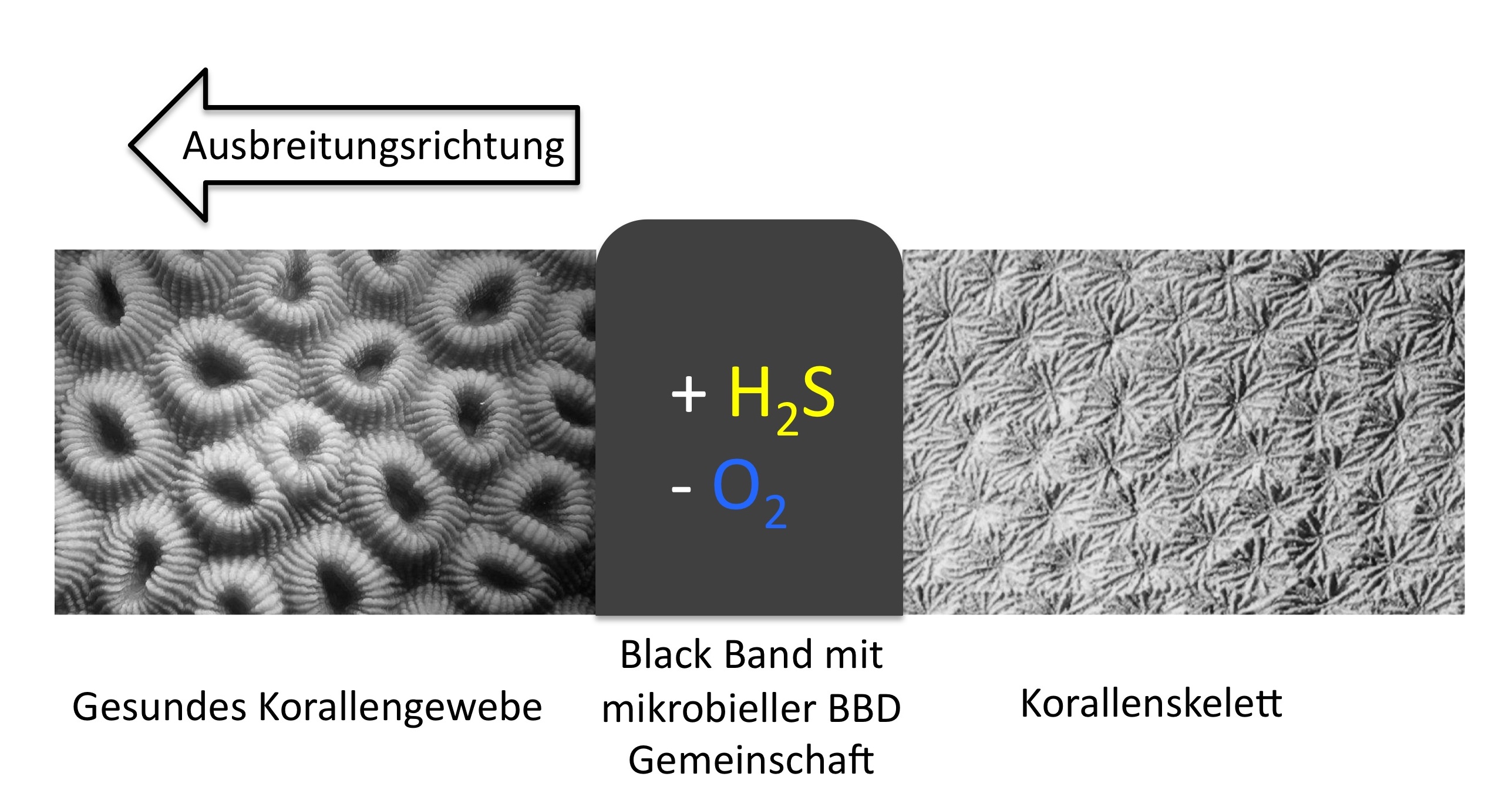
Figure 1: The dark front of the BBD migrates towards the healthy tissue and leaves the bare coral skeleton behind. Sulfidic (+H2S) and anoxic (-O2) conditions are responsible for the necrosis of the coral tissue in the BBD zone. Image: M. Glas/R. Dunker
The scientists investigated the tissue lesions with microsensors for oxygen, sulfide and pH. These microprobes have a tip diameter in the micrometer range and allow the scientists to measure highly resolved depth profiles in the coral tissue. They identified big differences between BBD infected tissue and tissue in the preliminary stage of the disease: “In diseased coral tissue two zones develop: A phototrophic zone at the top in which the cyanobacteria produce oxygen and a lower anoxic zone in which the bacteria degrade the necrotic coral tissue. Sulfide is formed in the degradation process”, Martin Glas explains the results (Figure 3). “In tissue that is only slightly infected the zonation is not nearly that strong. Usually we could not detect sulfide, and oxygen penetrated deep into the bacterial mat”.
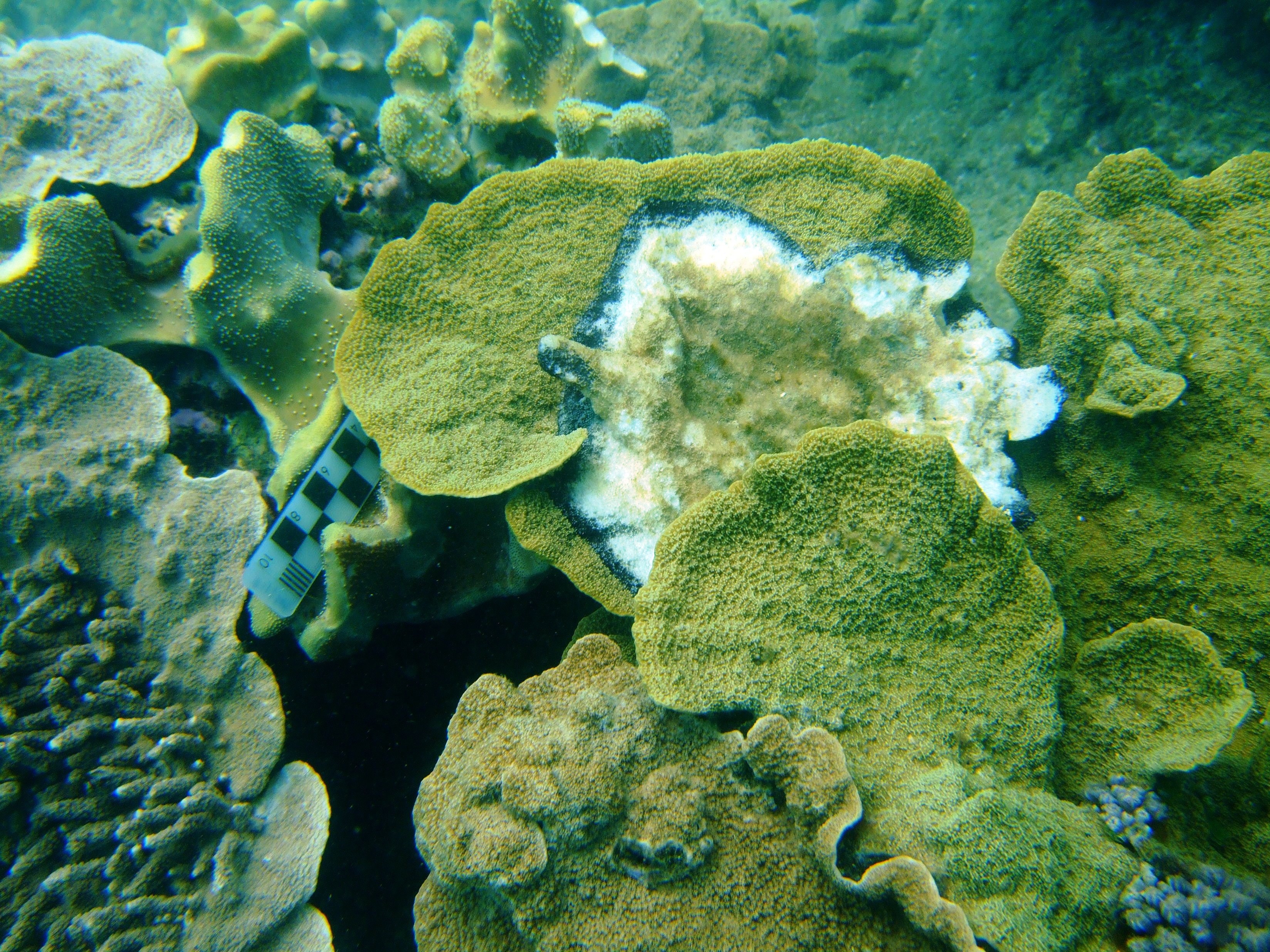
Figure 2: This coral in the Great Barrier Reef is strongly affected by the Black Band Disease. In the lower region the white limestone skeleton is visible, in the upper region the coral tissue is still intact. The BBD zone appears as a black band. Photo: Y. Sato
The corals and their endosymbiotic algae are struck by three stress factors at once: toxic sulfide, anoxia, and a low pH at the boundary of the bacterial mat and the coral tissue. At the front of the dark zone the conditions are particularly detrimental for the corals. The increased sulfide concentration around the necrosing tissue and the resulting decrease in oxygen leads to the spreading of the lesions to the surrounding, healthy tissue; a positive feedback that causes rapid migration of the BBD.
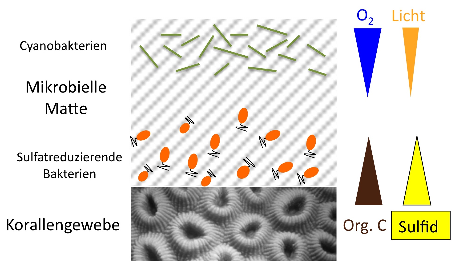
Figure 3: The section through the microbial mat on top of the coral tissue shows the incident light into the mat and the related oxygen production by the cyanobacteria. The lysing coral tissue releases organic carbon that is used by the sulfate reducing bacteria, and sulfide. The tissue lesion of the corals is thus a positive feedback process. Image: M Glas/R. Dunker
“We assume that the biogeochemical conditions at the surface of the coral tissue are responsible for the fast spreading of the disease. The higher the sulfide concentrations are and the less oxygen there is, the faster the dark front is migrating”, Martin Glas describes the causes for the origin and the high virulence of the BBD. So far, at least, the scientists have not identified a pathogen that could be responsible for the necrosis of the coral tissue.
For several years, David Bourne of the Australian Institute of Marine Science in Townsville and his colleague Yui Sato have been performing monitoring programs on the condition of coral reefs in which they also examined the coral diseases in the Great Barrier Reef. David Bourne says: „ Presumably the BBD is one of the most frequently reported diseases in tropical reefs. One major cause is the seasonally high water temperature. Thus, results from this study allow us to understand at the micro-scale how the environmental conditions and the complex microbial community interact to result in the onset and progression of this coral disease.”
Is there any cure for the reefs? “If the temperature decreases in winter the BBD is stagnant. However, with increasing frequency the disease recurs in the next year. The bare coral skeleton can be overgrown by new polyps. But this may take many years”, as Yui Sato of the James Cook University states.
Rita Dunker
For further information please contact
Martin Glas [Bitte aktivieren Sie Javascript]
Dr. David Bourne [Bitte aktivieren Sie Javascript]
Or the public relation office
Dr. Rita Dunker [Bitte aktivieren Sie Javascript]
Dr. Manfred Schlösser [Bitte aktivieren Sie Javascript]
Original article
Biogeochemical conditions determine virulence of black band disease in corals, 2012. M. S. Glas, Y. Sato, K. E. Ulstrup, and D. G. Bourne. The ISME Journal, advance online publication.
DOI: 10.1038/ismej.2012.2
Involved institutions
Max Planck Institut for Marine Micorbiology, Bremen
Centre of Microbiology and Genetics, Australian Institute of Marine Science, Townsville, Australia
ARC Centre of Excellence for Coral Reef Studies and School of Marine and Tropical Biology, James Cook University, Townsville, Australia
Australian Institute of Marine Science (AIMS), James Cook University, Townsville, Australia
DHI Water and Environment, West Perth, Australia