Seitenpfad:
- Abteilungen
- HGF MPG Brückengruppe für Tiefsee-Ökologie und -Technologie
- Planar Optode in situ module
Planar Optode in situ module
Die Inhalte dieser Seite sind leider nicht auf Deutsch verfügbar.
Frank Wenzhöfer
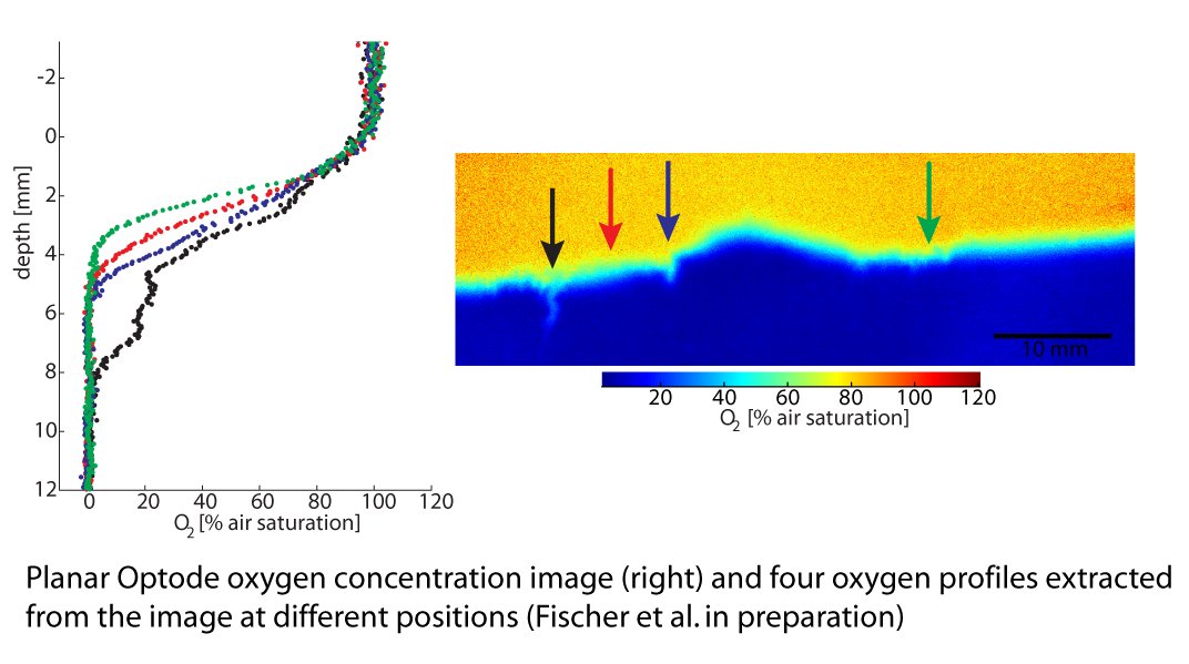
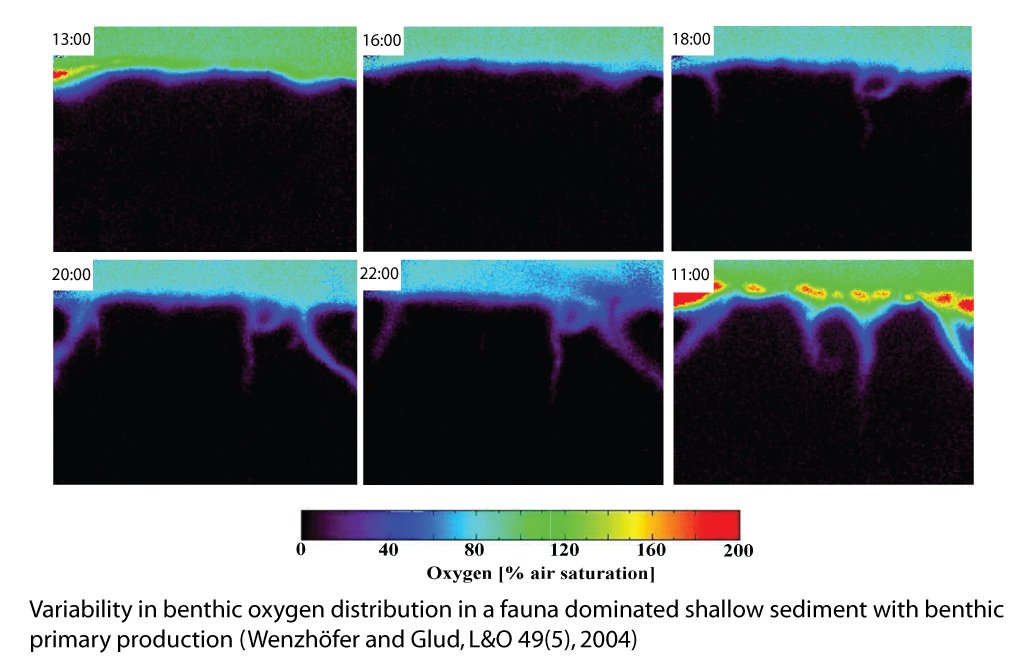
The distribution of oxygen in marine sediments is often highly heterogeneous due to factors like advection, bioturbation / bioirrigation, plant roots and benthic photosynthesis. The planar optode module enables in situ exploration of the oxygen dynamics at the sediment surface. The sediment oxygen distribution is recorded as 2D-images with a high temporal resolution giving insight to the spatial heterogeneity and its evolution over time.
The instrument
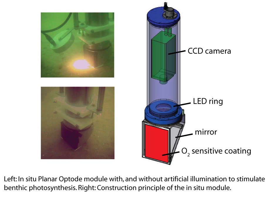
The in situ planar optode module consists of two pressure-proof and corrosion resistant titanium cylinders. One cylinder is equipped with a fast and sensitive CCD-camera, high power LEDs as a light source and an inverted periscope, which is inserted into the sediment allowing imaging of a cross section of the sediment. The second cylinder contains a computer which controls the camera and stores the images. The system, which is battery-powered, has a maximum operational time of 15 hours for continuous measurements and is operational even at full ocean depth (6000m).
For measurements of sediment oxygen distribution, the window of the inverted periscope is coated with a thin layer of oxygen sensitive fluorescent dye. The dye is illuminated with high power LEDs and the emitted florescent light is recorded as digital images by the camera. The intensity and lifetime of the emitted light, which can be calculated from the images, are dependent on the oxygen concentration at the respective position. In this way, each pixel of the digital images can be assigned an oxygen concentration value generating a high resolution 2D image of the oxygen distribution in the sediment.
Further improvements
Imaging of other analytes like pH and pCO2 are planned, as well as a reduction of size and power consumption of the whole system in order to increase the maneuverability and extend the long term applications of the planar optode module.
Since the core system is identical with the Multi Fiber Optode (MuFO), the technical improvements in either instrument can directly be transferred to the other
The instrument
The in situ planar optode module consists of two pressure-proof and corrosion resistant titanium cylinders. One cylinder is equipped with a fast and sensitive CCD-camera, high power LEDs as a light source and an inverted periscope, which is inserted into the sediment allowing imaging of a cross section of the sediment. The second cylinder contains a computer which controls the camera and stores the images. The system, which is battery-powered, has a maximum operational time of 15 hours for continuous measurements and is operational even at full ocean depth (6000m).
For measurements of sediment oxygen distribution, the window of the inverted periscope is coated with a thin layer of oxygen sensitive fluorescent dye. The dye is illuminated with high power LEDs and the emitted florescent light is recorded as digital images by the camera. The intensity and lifetime of the emitted light, which can be calculated from the images, are dependent on the oxygen concentration at the respective position. In this way, each pixel of the digital images can be assigned an oxygen concentration value generating a high resolution 2D image of the oxygen distribution in the sediment.
Further improvements
Imaging of other analytes like pH and pCO2 are planned, as well as a reduction of size and power consumption of the whole system in order to increase the maneuverability and extend the long term applications of the planar optode module.
Since the core system is identical with the Multi Fiber Optode (MuFO), the technical improvements in either instrument can directly be transferred to the other
Technical details
- Max. imaged field: 120 x 160mm
- Camera resolution: 1280 x 1024 pixels
- Max. spatial resolution: ~100µm
- Analytical range: 0 – 500µM oxygen
- Detection limit : 0.5µM oxygen
- Resolution: 0.1 µM oxygen
- Accuracy: 5%
- Response time: ~10s